the ear anatomy
Eye and Ear Models. 8 Pictures about Eye and Ear Models : Ear Cartilage | ClipArt ETC, The Radiology Assistant : Temporal bone - Anatomy 2.0 | Medical anatomy and also Tympanic membrane rupture - WikEM.
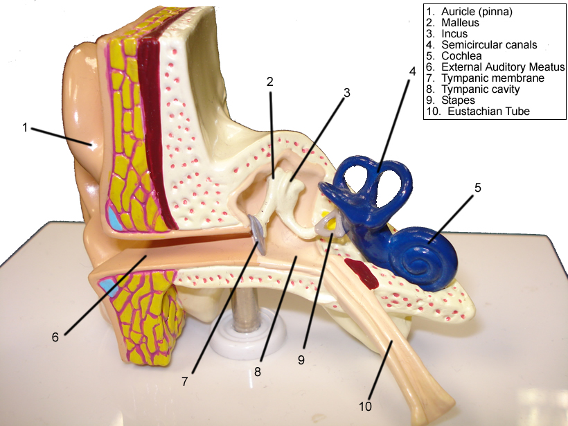
Eye And Ear Models
 www.biologycorner.com
ear anatomy eye models lab guide structures identify biologycorner brain senses biology eyes physiology parts answers
Parapharyngeal And Retropharyngeal Spaces: Anatomy | Kenhub
carotid artery common anatomy retropharyngeal parapharyngeal space spaces pharynx dorsal kenhub skull variations anatomical
Frontal Bone: Anatomy, Borders And Development | Kenhub
frontal bone kenhub anatomy frontale os
Medial Plantar Muscles Of The Foot: Anatomy | Kenhub
foot anatomy kenhub muscles plantar medial muscle ankle plantaris sole body diagram human sink 3d nerve study tibial anatomie read
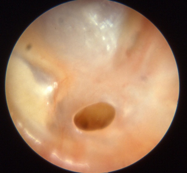
Tympanic Membrane Rupture - WikEM
 www.wikem.org
membran timpani perforasi tympanic membrane rupture perforation timpanica otitis clogged eardrum otite wikem ruptured plugged bacheca
The Radiology Assistant : Temporal Bone - Anatomy 2.0 | Medical Anatomy
 www.pinterest.com
anatomy temporal radiology eustachian istant dysfunction
Ear Cartilage | ClipArt ETC
ear cartilage cartilages etc clipart usf without edu medium
Normal Chest X-ray: Anatomy Tutorial | Kenhub
ray chest normal anatomy kenhub
Parapharyngeal and retropharyngeal spaces: anatomy. Normal chest x-ray: anatomy tutorial. Frontal bone kenhub anatomy frontale os