the anatomy of the eye
Figure 1: The right eye demonstrating the tear trough anatomy (top left. 9 Images about Figure 1: The right eye demonstrating the tear trough anatomy (top left : Anatomy – Brisbane Retina | Dr Abhishek Sharma, Figure 1: The right eye demonstrating the tear trough anatomy (top left and also Anatomy – Brisbane Retina | Dr Abhishek Sharma.
Figure 1: The Right Eye Demonstrating The Tear Trough Anatomy (top Left
 www.pinterest.com
trough orbicularis orbital inferomedial demonstrating
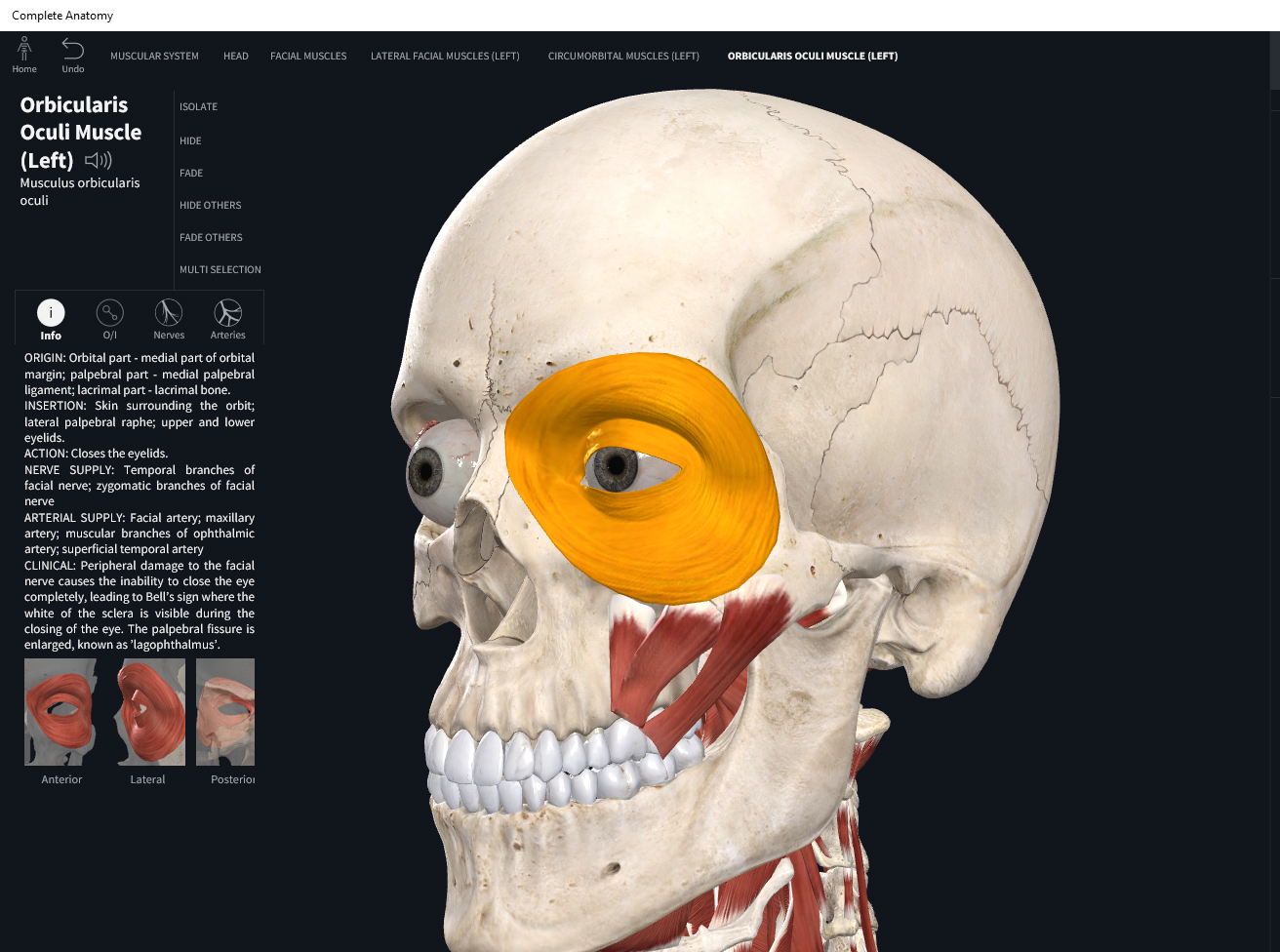
Muscles: Orbicularis Oculi. – Anatomy & Physiology
 integrativewellnessandmovement.com
orbicularis oculi
Third Eye Wallpapers (57+ Images)
 getwallpapers.com
third eye wallpapers
Anatomy Of The Cornea - TrialExhibits Inc.
 www.trialexhibitsinc.com
cornea
Untitled Document [bio.sunyorange.edu]
![Untitled Document [bio.sunyorange.edu]](http://bio.sunyorange.edu/updated2/comparative_anatomy/anat.html2/pignervous/n_eye.jpg) bio.sunyorange.edu
bio.sunyorange.edu
eye pig anatomy comparative
Dissected Cow Eye Clearly Showing How Eye Can Be Understood And How
 www.pinterest.com
cow lens anatomy eye dissected dissection labeled human eyes camera vet sagittal section understood clearly showing muscle open
20 Interesting Facts About The Eyes
 www.lolwot.com
eyes facts interesting
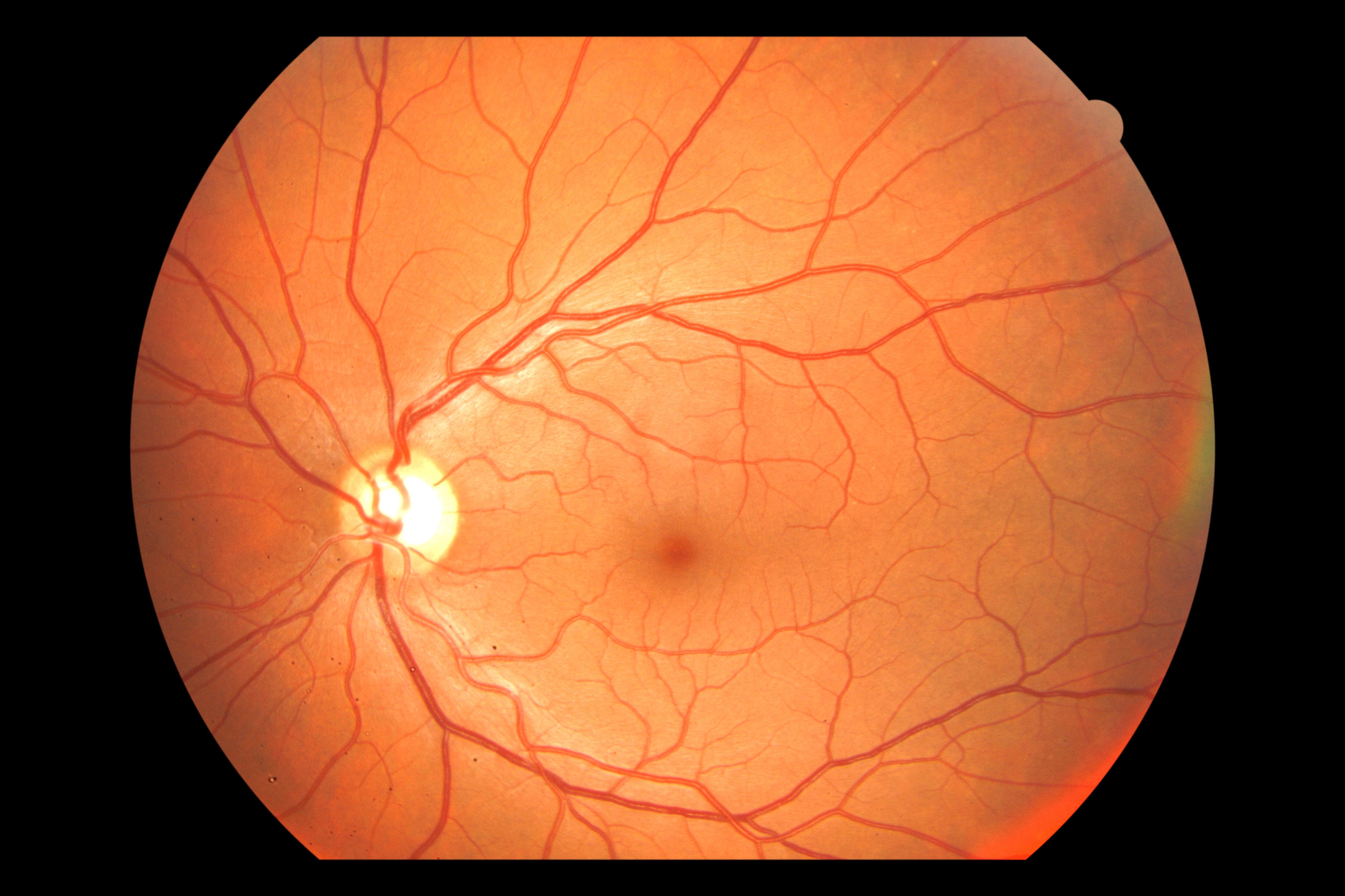
Anatomy – Brisbane Retina | Dr Abhishek Sharma
 brisbaneretina.com.au
retina normal anatomy fundus layers
Urinary Sys.: Kidney Model | Medical Anatomy, Human Body Anatomy
 www.pinterest.com
kidney anatomy human body
Retina normal anatomy fundus layers. Anatomy of the cornea. Eyes facts interesting