terminal ileum anatomy
87 Crohn Disease | Radiology Key. 8 Pics about 87 Crohn Disease | Radiology Key : Fat halo in normal terminal ileum | Radiology Case | Radiopaedia.org, Villous mucosa of the terminal ileum - YouTube and also Appendix | UW Ultrasound.
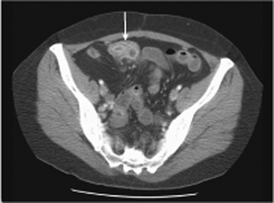
87 Crohn Disease | Radiology Key
 radiologykey.com
crohn disease ct fat terminal stranding ileocecal thickening cecum mild radiology axial junction inflammatory peri fig shows radiologykey
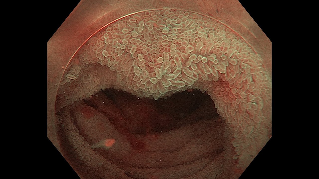
Villous Mucosa Of The Terminal Ileum - YouTube
 www.youtube.com
ileum terminal mucosa
Fat Halo In Normal Terminal Ileum | Radiology Case | Radiopaedia.org
 radiopaedia.org
terminal ileum normal halo fat ct radiopaedia case
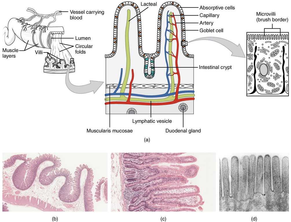
Difference Between Small And Large Intestine | Definition, Components
 pediaa.com
intestine difference between histology vs figure
Vascular Anatomy Of The Jejunum; Jejunal Root Artery, Arcade Artery
jejunum artery vasa recta jejunal vascular
Fat Halo In Normal Terminal Ileum | Image | Radiopaedia.org
 radiopaedia.org
fat ileum terminal normal halo infiltration radiopaedia ct version
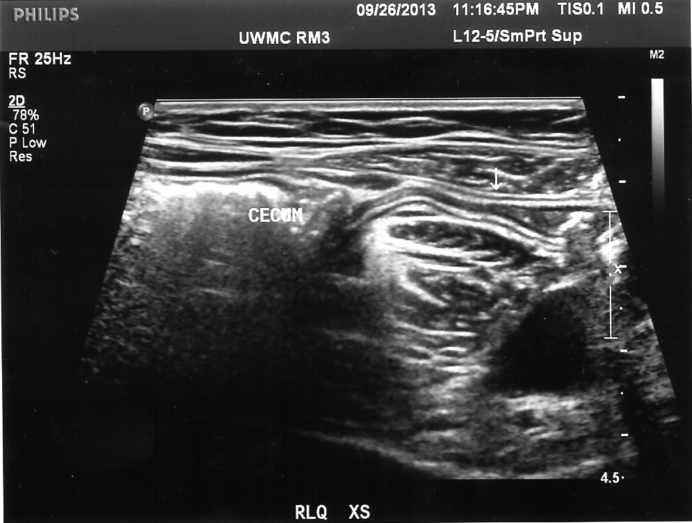
Appendix | UW Ultrasound
 depts.washington.edu
ultrasound cecum cecal joins
PPT - Anatomy Of The Kidney & Ureter PowerPoint Presentation - ID:2240729
 www.slideserve.com
ureter anatomy kidney anterior relation ppt duodenum powerpoint presentation
Intestine difference between histology vs figure. Terminal ileum normal halo fat ct radiopaedia case. Ultrasound cecum cecal joins