swallowing anatomy diagram
Normal laryngeal anatomy. Axial CT image at the level of the glottis. 9 Images about Normal laryngeal anatomy. Axial CT image at the level of the glottis : 17 Best images about Swallowing on Pinterest | Instrumental, Problem, Tracheostomy tubes - using a speaking valve fact sheet | Children’s and also Moth Anatomy - Anatomy Drawing Diagram.
Normal Laryngeal Anatomy. Axial CT Image At The Level Of The Glottis
 www.researchgate.net
anatomy glottis laryngeal axial cords laryngectomy commissures
The Basic Muscles In The Human Body | These Bones Of Mine
 thesebonesofmine.wordpress.com
muscles human body anterior labeled basic bones
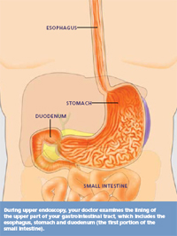
ASGE | Understanding Upper Endoscopy
 www.asge.org
endoscopy upper gi understanding egd patient asge illustration
Tracheostomy Tubes - Using A Speaking Valve Fact Sheet | Children’s
 www.childrens.health.qld.gov.au
speaking valve tracheostomy does tubes using fact health
Muscles And Tendons In The Hand – Art As Applied To Medicine
 medicalart.johnshopkins.edu
hand tendons muscles adobe weissbrod photoshop elizabeth illustration
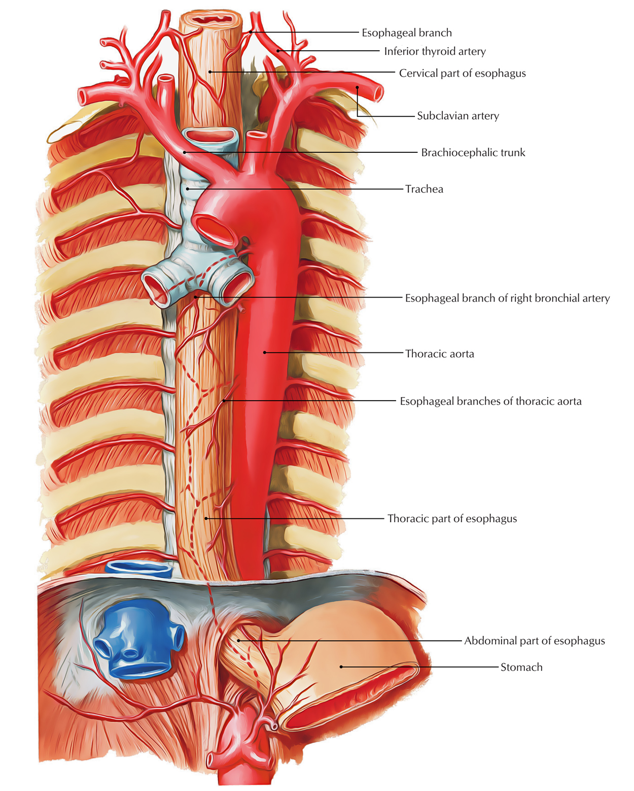
Easy Notes On 【Esophagus 】Learn In Just 4 Minutes! – Earth's Lab
 www.earthslab.com
esophagus aorta descending structure anatomy course parts earthslab its constrictions thorax arterial nerve venous column spinal left
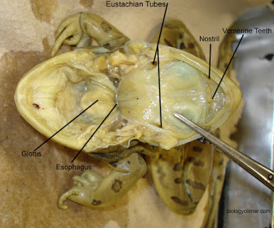
Frog External Anatomy
 www.biologycorner.com
frog dissection labeled mouth external tongue teeth anatomy glottis lab internal esophagus eustachian does structure found biologycorner pre attach study
Moth Anatomy - Anatomy Drawing Diagram
 sen842cova.blogspot.com
moth tiger cream anatomy moths spot arctia diagram wings garden caterpillar insight wildlife
17 Best Images About Swallowing On Pinterest | Instrumental, Problem
 www.pinterest.com
swallowing speech language swallow therapy dysphagia oral disorders pathology aging tools feeding motor handout adult swallows asha córtex cerebral activities
Frog dissection labeled mouth external tongue teeth anatomy glottis lab internal esophagus eustachian does structure found biologycorner pre attach study. 17 best images about swallowing on pinterest. Muscles human body anterior labeled basic bones