surface bone anatomy mri scan
Mandibular tori | Image | Radiopaedia.org. 9 Pics about Mandibular tori | Image | Radiopaedia.org : Radiological anatomy : X-ray, CT, MRI | Kenhub, Mandibular tori | Image | Radiopaedia.org and also A New Numerical Model to Analyze Stress Distribution of TMJ Disc from 2.
Mandibular Tori | Image | Radiopaedia.org
 radiopaedia.org
mandibular tori radiopaedia torus mandible radiology bone version
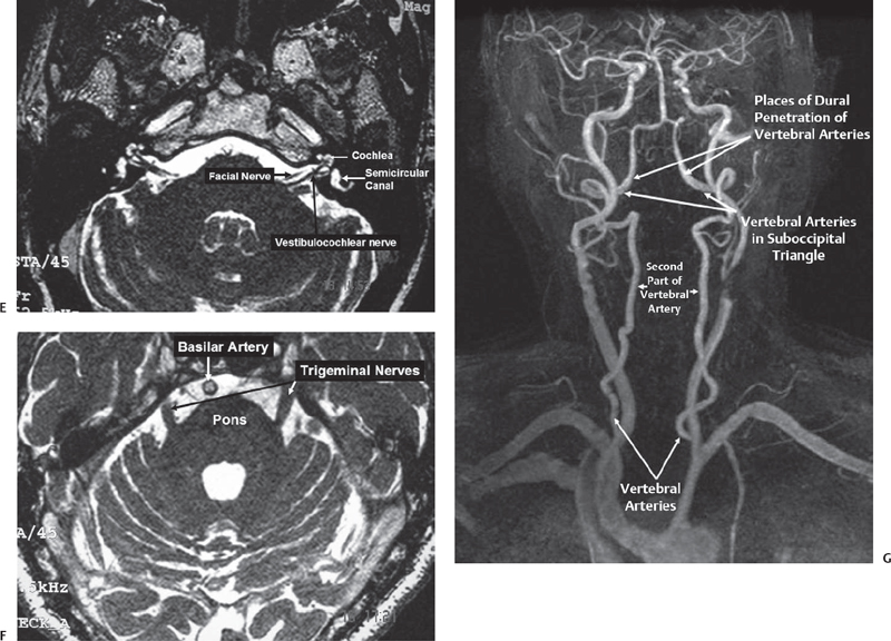
Microsurgical Anatomy Of The Posterior Cranial Fossa | Neupsy Key
 neupsykey.com
fossa posterior anatomy cranial mri sagittal microsurgical
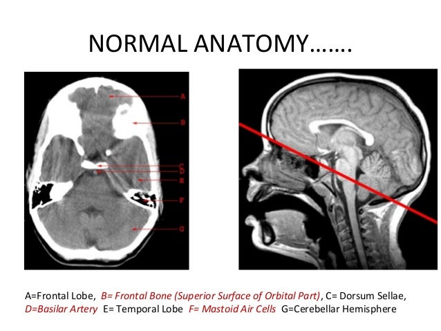
CT Scan Head Basics
 www.slideshare.net
lobe cerebellar temporal frontal
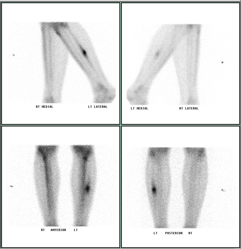
Nuclear Medicine Technology Program: Stress Fracture - Shin Splints
 umdnjnmt.blogspot.com
fracture bone stress shin splints nuclear medicine pain program technology subsides displacement symptom associated rest which
A New Numerical Model To Analyze Stress Distribution Of TMJ Disc From 2
 article.sapub.org
Xray Picture Brain Mri Scan Image Stock Photo 51730837 - Shutterstock
hersenen meningioma resonantie magnetische magnetisch gehirn resonanz sclerosis destra frontale resonance progression magnetica medisch mensen transverse slowed craniotomia risonanza ray
The 'beauty' Of The Head And Neck: From Temporal Bone To Oral Cavity
 www.auntminnie.com
neck midline floor head mouth dermoid cyst sagittal contrast cavity oral cystic lesion muscles enhanced weighted t1 coronal hourglass shaped
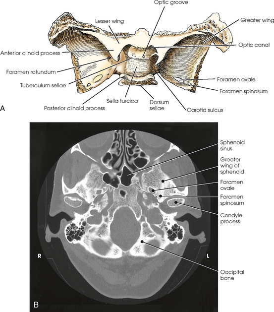
SKULL | Radiology Key
 radiologykey.com
sphenoid ct wing greater lesser skull bone radiology superior scan axial base wings lateral aspect slidesharetrick key radiologykey
Radiological Anatomy : X-ray, CT, MRI | Kenhub
mri caudate nucleus radiological imaging kenhub t2w csf
Fossa posterior anatomy cranial mri sagittal microsurgical. Neck midline floor head mouth dermoid cyst sagittal contrast cavity oral cystic lesion muscles enhanced weighted t1 coronal hourglass shaped. Hersenen meningioma resonantie magnetische magnetisch gehirn resonanz sclerosis destra frontale resonance progression magnetica medisch mensen transverse slowed craniotomia risonanza ray