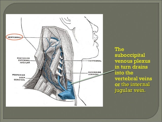
suboccipital venous plexus
Scalp. 9 Images about Scalp : Contribution of embryology in the understanding of cervical venous, Head and neck – MistryLand and also Surgical Anatomy of the Posterior Part of the Foramen Magnum and the.
Scalp
 www.slideshare.net
scalp
Contribution Of Embryology In The Understanding Of Cervical Venous
 link.springer.com
venous cervical transverse vein vertebral system fig loop 1206
Craniocervical Junction Venous Anatomy On Enhanced MR Images: The
 www.ajnr.org
suboccipital sinus cavernous venous craniocervical enhanced mr junction anatomy ajnr fig
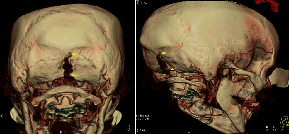
Cureus | Abnormal Large Central Occipital Emissary Vein: A Case Report
 www.cureus.com
occipital vein emissary figure abnormal literature central case report venous cta reconstruction plexus suboccipital showing course 3d cureus
Surgical Anatomy Of The Posterior Part Of The Foramen Magnum And The
 plasticsurgerykey.com
foramen magnum anatomy posterior surgical paramedian approaches structures veins dural fig craniotomy fig2
A Chiropractic Lifestyle: Is It Normal To Feel Lightheaded After An
 dramyreich.blogspot.com
muscles suboccipital skull neck cervical head upper atlas c1 feel nerve trigger triangle headaches normal themselves chiropractic lifestyle points plexus
Suboccipital Triangle Musculature | Neuroanatomy | The Neurosurgical Atlas
 www.neurosurgicalatlas.com
suboccipital musculature surgical neuroanatomy neurosurgicalatlas
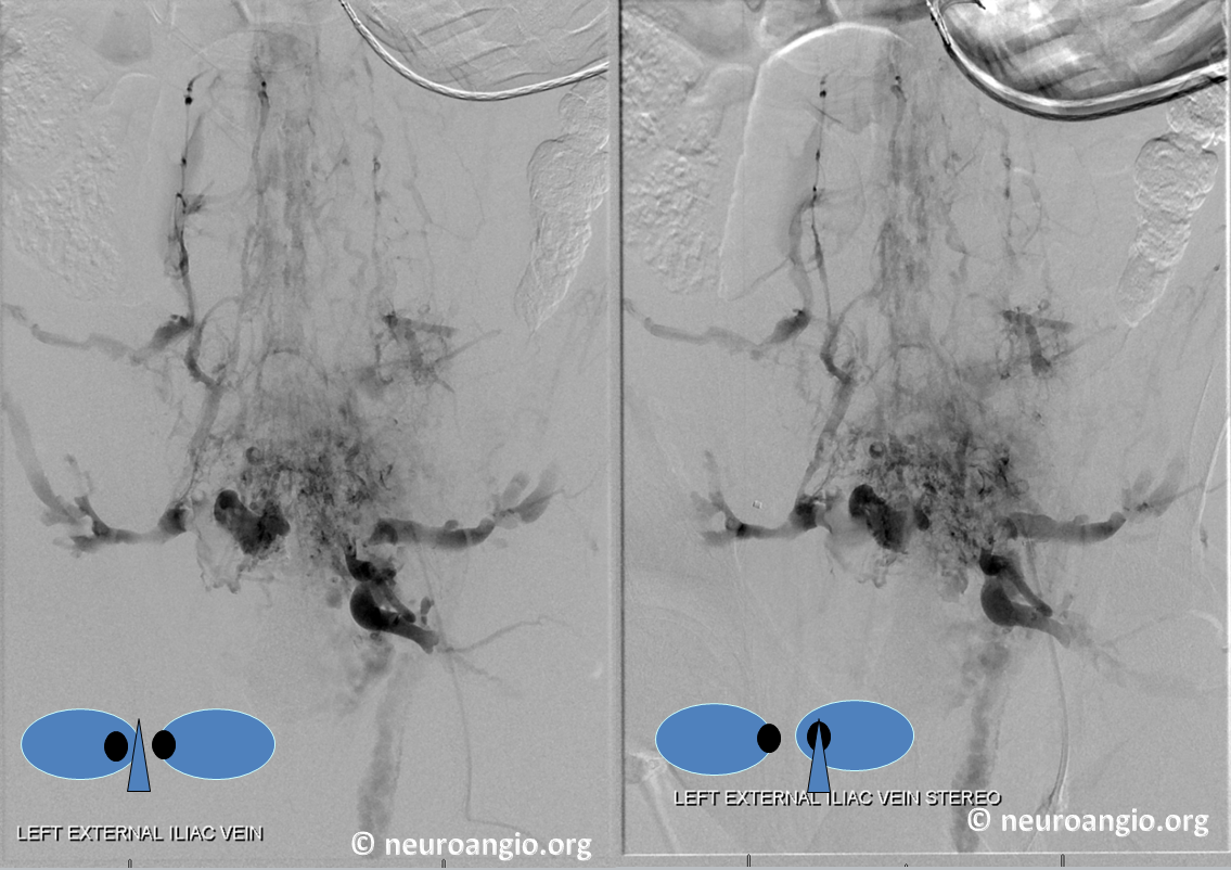
Archives Batson Venous Plexus | Neuroangio.org
 neuroangio.org
plexus batson venous archives neuroangio views lateral lumbar region
Head And Neck – MistryLand
 mistry07.wordpress.com
suboccipital muscles triangle artery neck skull vertebral base tension headache contents left head occipital capitis boundaries right attach deep attachments
Venous cervical transverse vein vertebral system fig loop 1206. Surgical anatomy of the posterior part of the foramen magnum and the. Foramen magnum anatomy posterior surgical paramedian approaches structures veins dural fig craniotomy fig2