spinal cord anatomy labeled
Sagittal MRI C-spine, T2, with structures labeled | Mri, Mri study. 9 Images about Sagittal MRI C-spine, T2, with structures labeled | Mri, Mri study : Nerve Models, The Spinal Cord, Nerve Roots and Cauda Equina - TrialExhibits Inc. and also Untitled Document [bio.sunyorange.edu].
Sagittal MRI C-spine, T2, With Structures Labeled | Mri, Mri Study
 www.pinterest.com
labelled mri spine sagittal labeled structures t2
Untitled Document [bio.sunyorange.edu]
![Untitled Document [bio.sunyorange.edu]](http://bio.sunyorange.edu/updated2/comparative_anatomy/anat_3/vertebrae_general/13a.jpg) bio.sunyorange.edu
bio.sunyorange.edu
notochord coelom lancelet lamprey amphioxus anatomy coelomate gonads hagfish cavity section cross heart worms cord frog rna hydra human sunyorange
Overview Of The Central Nervous System (Gross Anatomy Of The Brain) Part 3
 what-when-how.com
gross brainstem ventral cerebral pons nervous peduncle decussation pyramidal nerves cranial basilar crus cerebri rostral basal olivary nucleus cortex anatomi
Transverse Section Of The Spinal Cord | ClipArt ETC
spinal cord section transverse edu etc clipart tiff usf
The Spinal Cord, Nerve Roots And Cauda Equina - TrialExhibits Inc.
 www.trialexhibitsinc.com
equina cauda
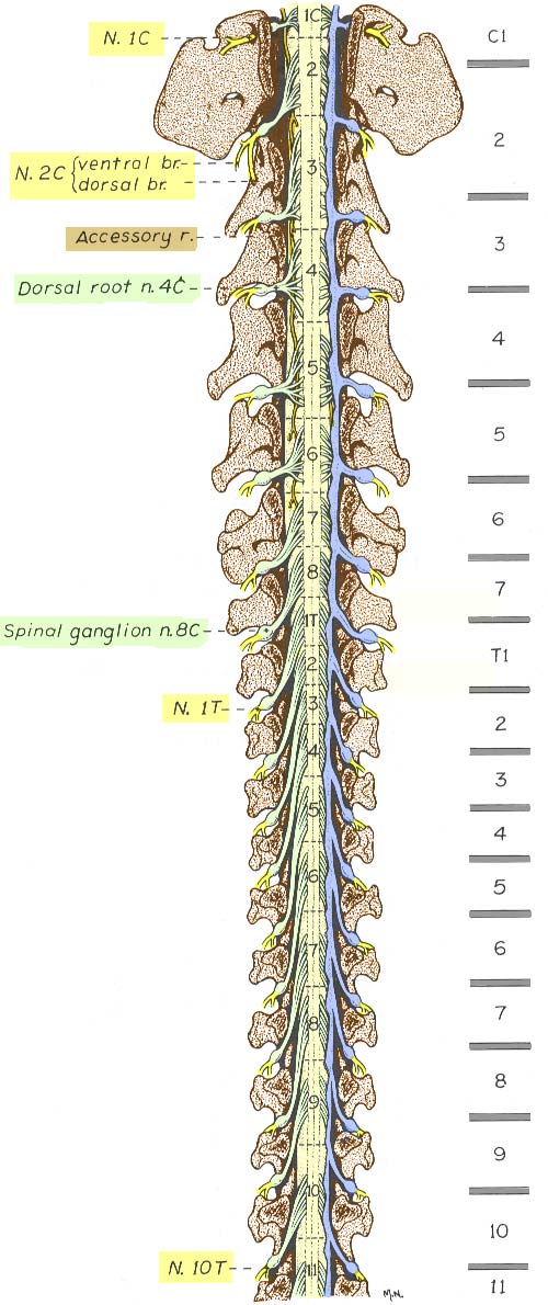
Spinal Cord Segments And Spinal Roots
 vanat.cvm.umn.edu
spinal cord column segments canine vertebral intervertebral labeled cranial thoracic mater dura plexus gross ganglia been roots disc bodies discs
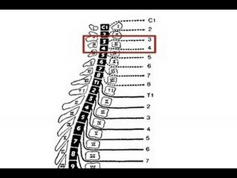
Levels Of Function In Spinal Cord Injury - YouTube
 www.youtube.com
spinal cord injury levels injuries therapy level function occupational physical medical nursing nurse neurology exam problems health different sci shock
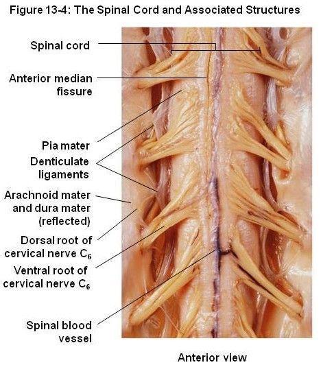
Print The Spinal Cord And Tracts Flashcards | Easy Notecards
 www.easynotecards.com
spinal cord mater denticulate ligaments pia dura anatomy human root dorsal ganglion structures column google identify anchor system easynotecards tracts
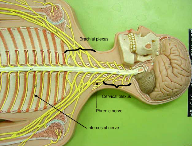
Nerve Models
 classroom.sdmesa.edu
nerve nerves anatomy models labeled plexus spinal nervous label system cervical lumbar brachial plexuses sacral human arm lab legs brain
Transverse section of the spinal cord. Gross brainstem ventral cerebral pons nervous peduncle decussation pyramidal nerves cranial basilar crus cerebri rostral basal olivary nucleus cortex anatomi. Sagittal mri c-spine, t2, with structures labeled