skeletal anatomy talus bone
Skeleton Atlas: The Complete Skeletal Anatomy: Skeletal System Images. 9 Pictures about Skeleton Atlas: The Complete Skeletal Anatomy: Skeletal System Images : Osteochondral Lesion of the Talus | Torn Ankle Cartilage, Anterior View Of Left Tarsal Bone And Ankle Diagram and also SKELETAL SYSTEM THE AUDITORY OSSICLES MEDIAL VIEW POSTERIOR VIEW.
Skeleton Atlas: The Complete Skeletal Anatomy: Skeletal System Images
 www.ebay.com
skeletal anatomy atlas system skeleton complete bone references fracture
SKELETAL SYSTEM THE AUDITORY OSSICLES MEDIAL VIEW POSTERIOR VIEW
 me.me
me.me
skeletal system anterior posterior lateral bone medial auditory ossicles happy bones process suture mandible parietal foramen
Osteochondral Lesion Of The Talus | Torn Ankle Cartilage
 www.hss.edu
talus ankle foot bones highlighted osteochondral lesion labeled illustration cartilage shown side
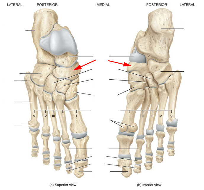
Skeletal Anatomy Of The Ankle And Foot Flashcards By ProProfs
 www.proprofs.com
anatomy foot ankle skeletal tibia fibula flashcards bone tarsal bones navicular called which proprofs proximal arch cuneiform talus cuboid intermediate
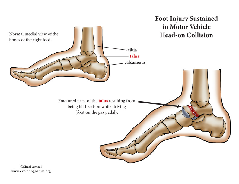
Ankle Injury (Talar Fracture) - Car Accident
 www.exploringnature.org
fracture talar accident talus
The Right Talus Bone Ex Situ | Anatomy Bones, Foot Anatomy, Ankle Anatomy
 www.pinterest.com
talus foot bone right anatomy navicular
Anterior View Of Left Tarsal Bone And Ankle Diagram
 www.anatomynote.com
ankle anatomy foot anterior diagram left bone ligaments medial tarsal human lateral bones sprain cuneiforms skeleton joint tendons leg shin
Bones Of The Lower Limb Page 6
 www.edoctoronline.com
lower bones limb limbs anatomy foot tibia fibula skeleton lateral trochanter medial intercondylar nonsense anatomical lesser talus atlas calcaneus condyles
Leg Bones - Medical Art Library
 medicalartlibrary.com
leg bones anatomy human skeleton medical lower bone foot knee joint muscles medicalartlibrary tibia lateral hip body legs ball end
Ankle injury (talar fracture). Bones of the lower limb page 6. Talus ankle foot bones highlighted osteochondral lesion labeled illustration cartilage shown side